24 X 7 online support mishr_c@hotmail.com
Medical Emergency +91 99242 31500
Menu

Severe Acute Inflammatory Demyelinating

 Severe Acute Inflammatory Demyelinating Polyneuropathy (AIDP): Comprehensive Critical Care, Family Partnership, and Early Rehabilitation Leading to Successful Recovery

A 71-year-old gentleman with hypertension, ischemic heart disease, prior coronary angioplasties, severe left ventricular dysfunction (ejection fraction 25%), a cardiac resynchronization therapy-defibrillator (CRT-D), and chronic treatment with beta-blockers, statins, dual antiplatelet therapy, and antihypertensive medications developed rapidly progressive acute inflammatory demyelinating polyneuropathy (AIDP).

Within 24 hours of symptom onset, he developed respiratory failure requiring endotracheal intubation and invasive mechanical ventilation. AIDP was subsequently confirmed, and intravenous immunoglobulin (IVIg) therapy was initiated before referral to our centre.

From our perspective, plasma exchange (plasmapheresis) would have been the preferred first-line immunotherapy for a patient with fulminant disease requiring mechanical ventilation. Although both IVIg and plasma exchange are evidence-based therapies, we often favour plasma exchange in such critically ill patients because treatment can be individualised, antibody removal is immediate, and clinical recovery may be facilitated in selected patients. However, because IVIg had already been initiated, the treatment course was completed.

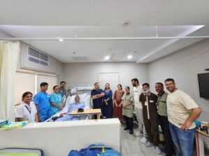
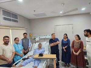
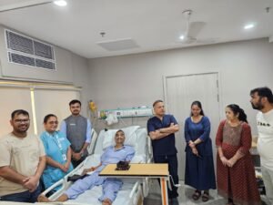
The patient remained awake, cooperative, and interactive throughout his ventilatory support. Sedation was deliberately minimized, with only low-dose fentanyl administered for comfort. Family members were encouraged to remain actively involved in patient care, providing emotional reassurance and continuous motivation, thereby greatly reducing anxiety and facilitating communication.

The ICU course was complicated by significant autonomic dysfunction, with fluctuating blood pressure and heart rate. Several cardiac medications were temporarily withheld, except aspirin, while low-molecular-weight heparin was continued for thromboprophylaxis. Careful haemodynamic monitoring and prompt management of autonomic instability allowed stabilization without major cardiovascular complications.

After approximately one week, ventilatory requirements had become minimal. However, repeated spontaneous breathing trials and two extubation attempts with high-flow nasal cannula support failed due to persistent bulbar weakness and an ineffective cough, despite adequate respiratory mechanics. Consequently, a tracheostomy was performed on day 10.

After tracheostomy, the patient was weaned from mechanical ventilation and maintained on high-flow oxygen via the tracheostomy. Early transfer from the ICU to the ward was possible because family members completed structured training in tracheostomy care, suctioning, positioning, nutrition, and recognition of clinical deterioration. This transition significantly shortened the ICU stay while maintaining patient safety.

Throughout the admission, the neurology team, under the guidance of Professor Sudhir Shah and colleagues, provided continuous expert input on disease progression and neurological

recovery. Their close collaboration with the critical care team enabled timely therapeutic decisions.

Strict antimicrobial stewardship was a key aspect of management. The patient repeatedly grew multidrug-resistant Acinetobacter species from respiratory samples and multidrug-resistant Klebsiella species from urine cultures. However, these findings represented colonization rather than active infection. In the absence of fever, hemodynamic instability, inflammatory response, or organ dysfunction suggestive of sepsis, antibiotics were intentionally withheld. This decision avoided unnecessary antimicrobial exposure and likely reduced the risk of further resistance.

Because neurological recovery remained slow after approximately three weeks, a second course of IVIg was initiated following a multidisciplinary discussion. On the fourth day of therapy, the patient developed septic shock. IVIg was immediately discontinued, appropriate antibiotics were started, and aggressive sepsis management was initiated without delay.

Remarkably, the patient’s family recognised the earliest signs of deterioration at approximately 4:00 a.m. and alerted the treating team immediately. Appropriate sepsis management was initiated within an hour. Early recognition and rapid intervention led to complete haemodynamic stabilisation within six hours, thereby avoiding ICU readmission. This episode highlighted the tremendous value of an educated and empowered family acting as an extension of the healthcare team.

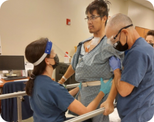
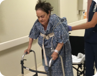
Subsequently, the tracheostomy tube was replaced with a speaking tracheostomy tube. Restoring speech significantly improved communication, emotional well-being, confidence, and active participation in rehabilitation. Simultaneously, aggressive multidisciplinary physiotherapy was intensified with advanced rehabilitation devices for upper- and lower-limb strengthening, progressive mobilisation, respiratory muscle training, and functional recovery. Nutritional support was carefully optimised throughout hospitalisation to preserve muscle mass and facilitate neurological recovery.

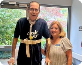
Progressive improvement in swallowing, respiratory function, and cough strength enabled successful decannulation within one week of switching to the speaking tracheostomy tube. During recovery, the patient was able to spend two to three days at home before returning for continued supervised rehabilitation, which substantially improved his confidence and psychological recovery.

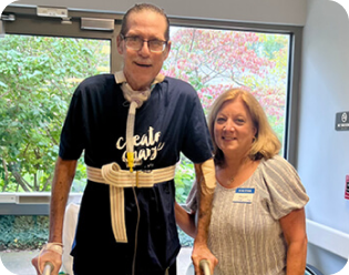
At discharge, the patient was breathing independently without a tracheostomy, able to speak normally, swallow liquids safely, walk with assistance, and actively participate in strengthening exercises. Distal muscle power had improved substantially, and proximal muscle recovery was progressing well.

Learning Points

This case demonstrates that successful recovery from severe ventilator-dependent AIDP requires far more than disease-specific immunotherapy. Recovery was driven by meticulous critical care, minimal sedation, management of autonomic dysfunction, judicious antimicrobial stewardship, structured family engagement, early tracheostomy, intensive

physiotherapy, optimized nutrition, multidisciplinary collaboration, and rapid recognition of complications.

Perhaps the most important lesson from this case is that when families are educated, empowered, and integrated into the healthcare team, they become active partners in recovery rather than passive observers. Such a comprehensive model of care has the potential to shorten ICU stays, reduce complications, improve functional outcomes, and help patients return to independent living much earlier than traditionally expected.

Dr. Rajesh Chandra Mishra, MBBS, MD, FNB (Critical Care Medicine), EDIC, FCCM, FCCP, FICCM, FICP

Senior Consultant – Intensive Care & Internal Medicine Sterling Hospitals, Ahmedabad, Gujarat, India

Director, Shaibya Comprehensive Care Clinic, Ahmedabad

Past President, Indian Society of Critical Care Medicine (ISCCM) (2022–2023)

Past General Secretary, ISCCM (2019–2020)

Past Chancellor, Indian College of Critical Care Medicine (ICCM)

Former India Representative to the European Society of Intensive Care Medicine (ESICM)

Editor-in-Chief of multiple textbooks in Critical Care Medicine, Extracorporeal Therapies, and Mechanical Ventilation

Author of numerous peer-reviewed publications, book chapters, national guidelines, and invited international lectures in Critical Care Medic

When Teamwork Makes the Impossible Possible

When Teamwork Makes the Impossible Possible

A Story of Physiology, Courage, and Multidisciplinary Critical Care

Introduction

Every intensivist encounters a few patients during his or her career who redefine the boundaries of what modern critical care can achieve. This was one such patient.

A 38-year-old gentleman sustained two close-range gunshot injuries, resulting in devastating thoracoabdominal trauma. One bullet traversed the abdomen, injuring the stomach, colon, and small intestine, narrowly missed the posterior walls of the aorta and inferior vena cava, and finally exited through the thorax, creating a large bronchopleural fistula. The extent of injury was catastrophic, involving multiple organ systems simultaneously and presenting one of the greatest challenges an intensivist can face.
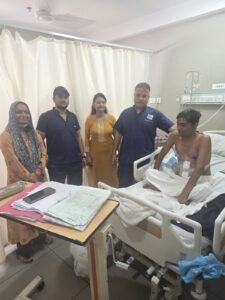
He underwent emergency damage-control surgery at the referring hospital. The primary objectives were haemorrhage control, contamination control, bullet removal, and stabilisation for transfer. An intercostal drain was inserted for the thoracic injury, and the patient was transferred to our tertiary care ICU approximately 24 hours later.

Although damage-control surgery had been successfully performed, the physiological battle was only just beginning.

Arrival at Our ICU: A Patient in Profound Physiological Collapse

When the patient arrived, he was critically ill and rapidly progressing towards irreversible multiple organ dysfunction.
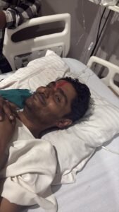
He remained hypotensive despite ongoing resuscitation, oliguric, and severely acidotic, with an arterial pH of 6.8, reflecting profound tissue hypoperfusion and ongoing shock. Liver function was severely impaired, with SGOT and SGPT exceeding 5,000 IU/L, suggesting ischemic hepatitis (“shock liver”). His coagulation profile was deranged after massive trauma and emergency surgery.

The respiratory challenge was equally formidable.

Due to the large bronchopleural fistula, more than 60% of each tidal volume delivered escaped through the chest drain, rendering conventional positive-pressure ventilation highly ineffective. Every breath delivered by the ventilator was partially lost through the fistula rather than contributing to alveolar gas exchange. Severe hypoxemia, hypercapnia, and ventilator instability became persistent threats.

At this stage, the patient had entered a vicious cycle:

 Persistent shock worsened tissue perfusion.

 Poor tissue perfusion aggravated metabolic acidosis.

 Acidosis impaired myocardial performance.

 Ongoing air leak prevented effective ventilation.

 Inadequate ventilation further worsened hypoxia and acidosis.

 Progressive inflammation increased the risk of irreversible multiorgan failure.

Without rapid physiological correction, survival was unlikely.

The First Decision: Treat Physiology Before Anatomy

Before proceeding with another surgical intervention, it was clear that the patient’s physiology had to be restored.

After detailed discussions with the family, thoracic and vascular surgeons, anesthesiologists, and the ICU team, we explained the extremely grave prognosis. The family showed extraordinary courage and trust. Despite understanding the significant risk of death, they encouraged us to pursue every reasonable intervention.
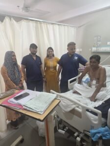
This unwavering support became one of the strongest pillars throughout the patient’s journey.

Our immediate objective during the next 48 hours was straightforward:

Restore physiology before attempting definitive surgery.

Advanced Hemodynamic Resuscitation

The cornerstone of management was individualised, physiology-guided resuscitation rather than protocol-driven fluid administration.

Advanced monitoring was established immediately:

 Invasive arterial blood pressure monitoring

 Central venous access

 Advanced cardiac output monitoring

 Serial bedside ultrasound examinations

 Continuous assessment of perfusion markers

 Frequent arterial blood gas analysis

Instead of administering large volumes of intravenous fluids empirically, every intervention was guided by dynamic physiological parameters. Repeated bedside ultrasonography senabled assessment of cardiac function, preload, ventricular filling, inferior vena cava dynamics, pulmonary congestion, and response to therapy. Cardiac output monitoring enabled continuous optimization of preload, afterload, and myocardial performance while avoiding both hypovolaemia and fluid overload.

This approach proved invaluable.

Despite severe shock, we successfully restored tissue perfusion without excessive crystalloid use.

One of our major goals was to avoid the “second hit” of fluid overload, increasingly recognised as an independent predictor of prolonged ventilation, abdominal compartment syndrome, renal dysfunction, delayed wound healing, and mortality in critically ill trauma patients.

Organ Rescue Rather Than Organ Replacement

Perhaps the greatest achievement during the first 48 hours was not merely stabilization but organ rescue. The kidneys had already begun to fail. The patient was oliguric and at extremely high risk of developing severe acute kidney injury requiring renal replacement therapy.

However, by restoring effective circulating volume, optimising cardiac output, maintaining adequate mean arterial pressure, correcting acidosis, and continuously reassessing organ perfusion, urine output gradually improved.
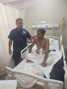
The patient never required dialysis.

Similarly, despite massive hepatic injury indicated by transaminase levels exceeding 5,000 IU/L, early restoration of oxygen delivery, correction of shock, and meticulous supportive care prevented progression to irreversible liver failure.

These outcomes reinforce a fundamental principle of critical care:

When tissue perfusion is restored early enough, injured organs often recover without requiring organ replacement therapy.

This concept—preserving organs before replacing them—remains one of the greatest strengths of modern intensive care.

Managing the Nearly Impossible: Ventilating a Massive Bronchopleural Fistula

The respiratory management of this patient posed one of the greatest technical challenges of the entire admission.

With over 60% tidal volume loss through the bronchopleural fistula, conventional ventilation became nearly ineffective. Each ventilator adjustment required careful physiological reasoning.

The objectives were simultaneously contradictory:

 Maintain adequate oxygenation.

 Minimize further air leak.

 Prevent ventilator-induced lung injury.

 Avoid worsening acidosis.

 Preserve hemodynamic stability.

Through individualised ventilator adjustments, meticulous monitoring, repeated blood gas analysis, and continuous bedside reassessment, we were able to maintain acceptable gas exchange despite the massive air leak. These 48 hours demanded constant vigilance from the ICU team.

Every small improvement represented another step toward definitive surgical repair.

By the end of this intensive physiological optimisation, the patient remained critically ill but had become stable enough to tolerate thoracic surgery. For the first time since his injury, survival appeared possible.

Source Control, Organ Support, and the Long Road to Recovery

The First Turning Point: Courage Beyond the Operating Room

After nearly 48 hours of meticulous physiological optimisation, the patient’s haemodynamics had begun to stabilise. Although he remained critically ill, there was a clear trend towards improvement. Lactate levels were declining, tissue perfusion had improved, urine output was recovering, and metabolic acidosis was slowly correcting. Yet one major obstacle remained—the massive bronchopleural fistula.

Despite all ventilatory adjustments, more than 60% of the delivered tidal volume continued to escape through the fistula. Effective ventilation remained almost impossible. It was clear that without definitive closure of the air leak, prolonged ventilatory support would become increasingly difficult, exposing the patient to worsening respiratory failure, ventilator-associated complications, and persistent physiological instability.

The decision to proceed with surgery at this stage required extraordinary courage.

Operating on a patient recovering from profound shock, severe metabolic acidosis, evolving multiorgan dysfunction, and major trauma is never routine. The risks of anaesthesia,

intraoperative instability, and perioperative mortality were substantial. However, delaying surgery posed an even greater risk.

At this crucial juncture, Dr. Bhupesh Shah Sir and his thoracic surgical team demonstrated remarkable clinical judgment and surgical courage. Recognising both the risks and the potential benefits, they proceeded with definitive repair of the bronchopleural fistula. The procedure marked the first major turning point in the patient’s recovery.

Immediately after fistula closure, ventilatory mechanics improved dramatically. The massive air leak disappeared, tidal volume delivery became effective, oxygenation stabilized, and ventilator settings could be gradually optimized. For the first time since admission,

respiratory support became manageable rather than merely salvage therapy.

This surgery fundamentally altered the trajectory of the patient’s illness.

A New Challenge: Persistent Sepsis Despite Physiological Improvement

Although respiratory mechanics improved significantly, the patient’s systemic inflammatory response remained unabated.

Over the following days, microbiological cultures revealed a succession of highly resistant pathogens:

 Multidrug-resistant Klebsiella

 Multidrug-resistant Acinetobacter

 Candida bloodstream infection (candidemia)

These findings reflected the complexity of penetrating abdominal trauma, emergency surgery,prolonged critical illness, and broad-spectrum antimicrobial exposure. Rather than relying solely on empirical escalation, antimicrobial therapy was continuously refined using culture reports, susceptibility testing, pharmacokinetic considerations, organ dysfunction, and daily multidisciplinary discussions involving microbiology, surgery, and critical care.

Antimicrobial stewardship became as important as antimicrobial therapy itself.

At the same time, meticulous supportive care continued:

 Daily assessment of fluid responsiveness

 Hemodynamic optimization

 Nutritional advancement

 Electrolyte correction

 Glycemic control

 Prevention of ICU-acquired weakness

 Prevention of secondary complications

Critical care is often described as “supporting the patient while nature heals.” In this patient, every hour required active intervention to preserve that opportunity for recovery.

Recognizing the Warning Signs

Despite intensive treatment, new concerns emerged.

The patient began experiencing episodes of unexplained hypoglycaemia, accompanied by renewed inflammatory markers and subtle clinical deterioration.

Rather than attributing these findings solely to ongoing sepsis, the ICU team considered the possibility of an uncontrolled intra-abdominal source.

A contrast-enhanced CT scan was urgently performed.

The imaging confirmed our concern.

There was evidence suggesting bowel leakage and ongoing intra-abdominal

contamination.

This represented another critical decision point.

Without definitive source control, no antibiotic regimen—regardless of its spectrum—would be sufficient.

The Second Turning Point: Surgery for Fecal Peritonitis

The patient was taken back to the operating room for emergency re-exploration.

Intraoperatively, Dr. Hitesh Chawada and his surgical team encountered severe faecal peritonitis due to gangrenous ischaemic transverse colon with ongoing contamination. Once again, surgery had to be performed on a patient who remained critically ill.

The operation involved resection of the nonviable colon, extensive peritoneal lavage, elimination of the septic focus, and creation of a colostomy.

This operation became the second major turning point in the patient’s recovery.

Source control remains one of the most important determinants of survival in abdominal sepsis. Even the most advanced intensive care cannot compensate for an uncontrolled septic focus.

By eliminating the source of infection, the physiological improvements achieved over the preceding days could finally be sustained.

Winning the Battle Against Organ Failure

Following definitive source control, the patient’s recovery accelerated.

Hemodynamic stability improved progressively.

Vasopressors were successfully discontinued.

Liver enzymes gradually normalized.

Renal function continued to recover without requiring renal replacement therapy, despite the severity of the initial acute kidney injury.

One particularly satisfying achievement throughout the patient’s ICU course was the judicious use of blood products.

Major trauma patients frequently receive large-volume transfusions, which may contribute to transfusion-associated lung injury, immunomodulation, volume overload, and infectious complications.

Through careful hemodynamic monitoring, restrictive transfusion practices, meticulous surgical hemostasis, and physiology-guided resuscitation, we avoided unnecessary transfusion.

During the entire ICU stay, the patient required only two units of packed red blood cells and two units of fresh frozen plasma.

Avoiding excessive blood product administration likely contributed to reduced pulmonary complications, improved immune function, and faster recovery.

Nutrition: An Often Underestimated Therapy

Critical illness is profoundly catabolic.

From the earliest feasible opportunity, nutritional therapy became a major component of

management.

Protein delivery was optimized.

Caloric targets were progressively achieved.

Micronutrient deficiencies were corrected.

Nutrition was continuously adjusted according to gastrointestinal function, metabolic demand, organ function, and surgical recovery.

Rather than viewing nutrition as supportive care alone, it was regarded as an active therapeutic intervention essential for wound healing, immune recovery, respiratory muscle strength, and successful liberation from mechanical ventilation.

Early Mobilization and Liberation from Mechanical Ventilation

As organ function improved, attention shifted toward recovery.

Our physiotherapy and rehabilitation team played an extraordinary role during this phase.

Passive mobilization began early.

Respiratory physiotherapy was intensified.

Muscle preservation became a priority.

Progressive mobilization minimized ICU-acquired weakness and prepared the patient for ventilator liberation.

Because respiratory mechanics had improved following fistula closure and sepsis was brought under control after abdominal surgery, the patient gradually fulfilled criteria for ventilator weaning.

Through careful physiological assessment, structured spontaneous breathing trials, and close multidisciplinary supervision, the patient was successfully extubated without requiring tracheostomy.

Following extubation, he required temporary support with high-flow nasal cannula (HFNC) before gradually transitioning to spontaneous breathing.

Avoiding tracheostomy in such a complex patient represented another important milestone in his recovery.

Recovery Beyond Medicine

Perhaps the most remarkable aspect of this patient’s journey was not only the science of critical care but also the humanity surrounding it.

Throughout these 26 days, the patient’s wife remained an unwavering source of strength.From the moment the patient became responsive, we encouraged structured family presence at the bedside. Their reassurance, emotional support, and constant encouragement became integral to rehabilitation.

Critical care extends beyond technology.

Healing also occurs through human connection.

In this patient, family involvement undoubtedly contributed to reduced anxiety, improved cooperation during weaning, enhanced motivation during rehabilitation, and ultimately a smoother recovery.

After 26 days of relentless multidisciplinary critical care, multiple surgeries, prolonged mechanical ventilation, severe sepsis, candidemia, multidrug-resistant infections, and repeated life-threatening complications, this young man walked out of the hospital with his family.

What initially appeared almost impossible had become reality—not because of one intervention, but because an entire team refused to give up.

Lessons Learned: What This Patient Taught Us

Every critically ill patient teaches us something. Some reinforce established principles, while a few redefine our approach to intensive care. This young man’s journey reaffirmed several fundamental concepts that every intensivist should keep in mind.

Physiology Before Procedures

The first and perhaps most important lesson was that physiology must be restored before

definitive intervention whenever possible.

Although the patient had a massive bronchopleural fistula requiring urgent surgical repair, immediate surgery in profound shock, severe metabolic acidosis (pH 6.8), evolving renal failure, and ischemic hepatitis would likely have carried an even higher mortality.

The initial 48 hours were therefore devoted to restoring physiology:

 Optimization of tissue perfusion.

 Goal-directed hemodynamic resuscitation.

 Cardiac output-guided fluid therapy.

 Bedside ultrasound-guided decision making.

 Careful ventilator optimization.

 Correction of severe metabolic derangements.

Only after achieving physiological stabilization was definitive surgery undertaken. This approach transformed an almost unsurvivable situation into one with a realistic possibility of recovery.

Monitoring Does Not Save Lives—Correct Interpretation Does

Modern intensive care provides access to sophisticated monitoring systems, but monitors themselves do not improve outcomes.

The correct interpretation of physiological data and timely therapeutic decisions are what save lives.

Repeated bedside ultrasound examinations, invasive arterial pressure monitoring, cardiac output monitoring, serial arterial blood gas analysis, and continuous reassessment enabled individualized management throughout the patient’s ICU stay.

Rather than administering empirical fluids or vasopressors, each intervention was guided by objective physiological evidence.

This individualized approach helped us restore perfusion while avoiding fluid overload.

Organ Rescue Is Better Than Organ Replacement

One of the greatest achievements in this case was preventing progression to irreversible organ failure.

Despite severe shock:

 The kidneys recovered without requiring renal replacement therapy.

 Severe ischemic hepatitis gradually resolved.

 Progressive multiorgan dysfunction was halted before irreversible damage occurred.

Critical care should always strive to preserve native organ function whenever possible. Organ replacement therapies remain invaluable rescue tools, but preventing their need through timely physiological optimization represents an even greater success.

Less Can Sometimes Be More

Trauma patients frequently receive large volumes of intravenous fluids and blood products. Current evidence increasingly supports balanced resuscitation while avoiding unnecessary transfusion and excessive positive fluid balance whenever clinically appropriate. Using advanced hemodynamic monitoring, dynamic assessment of fluid responsiveness, serial ultrasound, and careful clinical judgment, we avoided indiscriminate fluid administration.

During the entire ICU course, the patient required only two units of packed red blood cells and two units of fresh frozen plasma.

Avoiding excessive transfusion likely contributed to better pulmonary recovery, reduced inflammatory burden, and earlier liberation from mechanical ventilation.

Source Control Cannot Be Replaced by Antibiotics

Despite broad-spectrum antimicrobial therapy and intensive supportive care, the patient continued to deteriorate until the ischemic colon and fecal peritonitis were identified and surgically treated.

This case once again demonstrated one of the most important principles in critical care:

Antibiotics support recovery. Source control enables recovery.

The timely surgical intervention by Dr. Hitesh Chawada and his team fundamentally changed the patient’s trajectory.

Nutrition, Rehabilitation, and Family Are Therapeutic Interventions

Critical care does not end with stabilization.

Recovery depends equally upon:

 Early nutritional optimization.

 Prevention of ICU-acquired weakness.

 Early physiotherapy.

 Respiratory rehabilitation.

 Psychological support.

 Family involvement.

The decision to encourage family presence once the patient became responsive proved invaluable.

His wife remained beside him throughout this journey. Her encouragement became part of the therapeutic process itself. Similarly, the extraordinary efforts of the physiotherapy team enabled progressive mobilization, successful ventilator weaning, avoidance of tracheostomy, and eventual functional recovery.

The True Heroes

Although this article describes physiology and clinical decision-making, this patient’s recovery belongs to an extraordinary multidisciplinary team.

My deepest appreciation goes to Dr. Bhupesh Shah Sir and his thoracic surgical team, whose courage in repairing the bronchopleural fistula under such hostile physiological conditions created the first opportunity for recovery.

My sincere gratitude to Dr. Hitesh Chawada and his surgical team for their timely recognition and decisive management of ischemic colon and faecal peritonitis, which achieved definitive source control and saved this patient’s life.

I extend my heartfelt thanks to the Respiratory Care Unit team, whose seamless coordination and safe transfer ensured continuity of critical care from the referring hospital to our ICU.

A special word of appreciation goes to Kaushal and Abhi, whose professionalism and dedication during transport were instrumental in this patient’s successful transfer.

I am immensely proud of my entire ICU team, whose commitment never wavered during these 26 demanding days.

A special note of gratitude goes to Vaishali, Nidhi, and Sachin for their tireless bedside care, meticulous monitoring, unwavering vigilance, and compassionate support. Their dedication exemplified the very best of intensive care nursing and contributed immensely to this patient’s recovery.

My sincere thanks also go to our respiratory therapists, physiotherapists, microbiology team, dietitians, technicians, and every member of the multidisciplinary ICU team. Each played a vital role in transforming critical illness into recovery.

Finally, I wish to acknowledge the patient’s wife and family.

Throughout every discussion, every difficult decision, and every setback, they demonstrated remarkable courage, trust, patience, and resilience. Their faith in the treating team inspired us to continue fighting when the outcome appeared uncertain.

Final Reflection

Critical care medicine is often perceived as a specialty dominated by technology.

In reality, technology is only one component.

Patients survive because physiology is understood, decisions are timely, surgeons are courageous, nurses remain vigilant, physiotherapists mobilize relentlessly, microbiologists guide therapy, families continue to believe, and multidisciplinary teams refuse to give up.

This patient reminded all of us that impossible outcomes are rarely achieved by extraordinary individuals; they are achieved by ordinary professionals working together with extraordinary commitment.

Twenty-six days earlier, survival appeared almost impossible.

Today, he walked out of the hospital with his family.

There can be no greater reward for an intensivist.

With warm regards and heartfelt gratitude,

Dr. Rajesh Chandra Mishra

MBBS, MD (Medicine), FNB (Critical Care Medicine), EDIC, FCCM, FCCP, FICCM, FICP

Senior Consultant – Intensivist & Internist -Sterling Hospitals, Ahmedabad

Director – Shaibya Comprehensive Care Ahmedabad, Gujarat, India

Past President, Indian Society of Critical Care Medicine (ISCCM) (2022–2023)

Past General Secretary, Indian Society of Critical Care Medicine (ISCCM) (2019–2020)

Past Chancellor, Indian College of Critical Care Medicine (ICCM) (2022–2023)

Former India Representative, European Society of Intensive Care Medicine (ESICM)

“Critical care is not merely about supporting failing organs—it is about restoring life

through physiology, teamwork, compassion, and the courage to persevere when hope appears

distant.”—RCM ..

demo-2

A 35-year-old male presents with severe burning neuropathic-type pain in both lower limbs for the past 6 months, significantly affecting sleep and daily function. Pain is continuous, resistant to pregabalin/gabapentin/duloxetine, and NCS + EMG + MRI + B12 and autoimmune/heavy metal/metabolic workup are normal.

Initially evaluated as possible small fibre neuropathy (SFN) or even functional pain, as no demonstrable abnormalities were detected. Psychiatry trial also failed.

Important new development:
Patient has been taking proton pump inhibitors (PPI) daily for >1 year due to chronic epigastric burning. Upper GI endoscopy revealed severe gastritis with Helicobacter pylori infection. He was started on standard H. pylori eradication triple therapy — and his epigastric pain resolved within 15 days.

This now brings attention to the recognized but under-discussed association between chronic H. pylori infection and unexplained sensory neuropathic pain, possibly due to:

  • Chronic immune activation → small fibre inflammation
  • Molecular mimicry / autoantibody-driven neuropathy
  • Increased pro-inflammatory cytokines (IL-6/TNF-α) affecting dorsal root ganglia
  • Subtle micronutrient malabsorption despite normal B12 level
  • Central sensitization triggered by chronic gut–brain axis stimulation

This opens the possibility that H. pylori–induced neuroinflammation or immune-mediated small fibre dysfunction could have been a contributor or amplifier of his neuropathic pain.

– Case 12

21 Year young male, went to his physician with compliant DOE and palpitation. Pateint was send to Cardilologist for ECHO. ECHO finding were suggestive of Pulmonary embolism , RA RV Dilated with normal LV function.Immediate CTPA- WAS done which confirmed PE. Pateint was given 18 mg reteplase bolus. At that time ,HR130, BP 130/80, SPO2 85% (RA). blood reports came after reteplase was given which showed platelet count 14,000, and HB 10GM%. REST of the routine reports were normal. Now patient was shifted under my care after these report came. when patient came to me His Heart rate 132, BP 120/70, SPO282%( RA) but patient is breathing comfortably ( RR-24).

This patient fall in category of chronic sub massive ( moderate PE). As he was not in shock, not acidotic, not in perarrest condition, not severe RV Dysfuction. Evidence do not support use of thrombolytics in MODERATE PE( clinicaly stable patient. Thogh thrombocytopenia report came later but retrospectively it look the pateint was just lucky that he did not bleed with such a low platelet count.

As he was shifted to my care immedietely post thromolysis ,there was neither any clinical improvementt or deterioration.

Post thromolytic ECHO showed RA/RV Dilated , PASP (50 ), WITH NORMAL LV function.

when patient came to me His Heart rate 132, BP 120/70, SPO282%( RA) but patient is breathing comfortably ( RR-24).

Patient responded to Oxygen ( 15litre NRVM) ,SPO2 99%, As he was non acidotic , and hemodynamicaly stable , we planned to manage further with LMWH . Workup sarted for thrombocytopenia and thrombophilia. He was found to have (APLA & ACLA) postive with ADsDNA postive . Steroid and HCQs was strtaed for same . His platelet stated improving after 24 hour. He was started on warf after 48 hours. It look 7 days to achieve INR OF 3.5 Then sropped LMWH. Platelets came to normal after 5 days.

With retrospective history ,he gave history of Cough and DOE since last 2 months.

We have discharged this patient on warf ( target INR :3.75-4.5), HCQS, WYSOLONE & Mycophenilate mofetil.

At discahge his HR-90, SPO2 (92%-94% ) on RA, BP 120/80, RR 18. PASP 40.

I am watchful , but it way be possible this Chronic , submassive PE patient may need surgical thrombectomy . I am not sure when I will advise for that . I believe to give 3 month trail of controlling SLE, AND MAITAINING INR of (3.75 to 4.5).

– Case 11

The 74 year, male, tobacco chewer, veterinary Doctor was under treatment for diabetes and hypertension since last 5 years by a diabetologist. Four months ago his sugar control went out of control. 24-hour sugar study was done by diabetologist blood reports. As per the reports has baseline sugar fluctuations has increased, HbA1c 7.5%, ESR 75. As per his diabetologist opinion, he was started on three OHA -(GLICLAZIDE, METFORMIN, SITAGLIPTIN AND ONE DOSE INSULIN TRESIBA 8 UNITS.)

As the patient was known, so came to me with reports for casual second opinion at the same time 4 month ago for getting endorsed that all is well. I wish to get some more reports which his diabetologist said no, saying that all these changes are due to uncontrolled diabetes and once diabetes gets under control all the reports will be ok.

So I wanted to find out the reason for high ESR and fluctuating uncontrolled sugar. TB and malignancy were high in my card so I ask them to get a screening PET -CT has done. The request was turned down by diabetologist and he was very particular in saying that let us wait till sugar in under control and ESR will come down and ESR is a non-specific test.

Then two month ago, on three antidiabetic drugs and tresiba his sugar got under control, HbA1c =6.5 and ESR CAME DOWN TO 45. I was still persistent for PET CT- Request declined.

A week ago a patient came with 5 kg weight loss, SGPT 95, ALK PHOSPHATASE 154, BILIRUBIN 1.8. ESR again rose to 74. ( All these reports patient was getting it done himself -in view of routine checks)

I got his CA-19-9( 38- high normal) MDCT abdomen and MRCP done, which showed an inoperable CA Pancreas. Endoscopic USG biopsy showed high-grade Adenocarcinoma.
Lesson – 1-High ESR in diabetes is not a normal finding.

2-Whenever sugar goes out of control in well-managed diabetes find the treatable causes and precipitating factors rather adding three or four drugs.

– Case 2 – Backache?

This 43 yr female, from affluent class, came to for second opinion 5month ago. She was suffering from bachache since last two month. No fever,no loss of apetite, no weight loss and pain was localised to lower back. She was seen by neurologist, orthopedics, rheumatologist, spine surgeons & OB&G. CBC,CT & MRI were normal. ESR 35.Reason for my opinoin was non relieving pain by all measures and she was advised ovarian cyst surgery for pain. As I was I was not convinced with small folicular cyst and pain relation I asked to go to other OB&G, where he said this may due to LS joint TB. As pateint has local tenderness. He send patient back to me. She had local tenderness. I got her montoux, which was positive (20*20). Usg neck abdomen normal. Repeat ESR-40. CRP 32.

I was of the opinion to start AKT. But patient wanted confirmation, so I advised PET-CT, which showed increased uptake in LS Joint, pancreas and small lesion at Right lung apex area. To get bug and culture it I advised to go LS joint biopsy and cs or navigation-MR guided needle aspiration at clevland USA. Patient went to spine surgeon for biposy where he not only advised against biposy but said it is nothing . Got HLADR27, brucella and ANA profile which was negative. He advised patient to go to ID. As patient was from very affluent class well to lots of supersepecialst ,they took opinion from many concerned superspecialist and everyone advised against AKT.
She was put on NEOCOXIA AND MINOCYCLINE THINKING that it is PID and even PET CT , MONTOUX , ESR AND CRP ALL WERE IGNORED. She GOT SYMPTOMATICALLY BETTER.

After 6 month they came back to me with more sevre pain, fever low grade and incresed CRP ,ESR. This time same ID person advised that she should be given trail. Repeat HRCT -THORAX ,increased lung invlovement, medustinal LN positive. Now last 15 day she is on full AKT.

Though I persued this patient so much and was so confident for dx which I could see well before time, but was not endorsed by other seniors so treatment got delayed for 6 moth only.

– Anasarca – Interesting Case 1

This 33 yr female was referred to me for generalised anasarca for 2 years. She has gained 50 kg weight and now was not able to do routine activity due to breathlessness on exertion. O/E Pulse 70/min, BP 170/100, SPO2 95%(RA), generalised pitting edema ( from face to toe). She was comfortable in lying position. CXR-B/L blunt CP ANGLE . ECHO -WNL, grade -2,diastolic dysfunction. She was being treated for
hypothyroidism (50mcg eltroxin), hypertension ( amlodipine-10 mg), dytor 40 mg tds . Her albumin level was 3.5 gm%.

So as suggested by most of you we did send CBC-N,LFT-N,RFT-N, Na118, K 4.2,URINE-R/M,(Albumin nil,-3-4 cells, No RBCs).Albumin 3.5
gm% total protein 6.5gm%. ECHO-N, USG -Normal kidney- B/L Pleural effusion, free fluid in abdominal wall and 3rd spaces. TSH > 100, FREE
T4- LOW, Random cortisol low. Antithyroid Antibody positive very high. Anti -TPO antibody positive.

Based on above finding our dx was autoimmune thyroiditis with severe hypothyroidism anad hypocortisolism. We 1st gave 100 mg
hydrocortisone and 300 MCG eltroxin. Next day we have started on 200mcg eltroxin, wysolone 10mg ,CILINDAPINE, RAMIRIL AND
TEORSEMIDE . She was send home and came back after 14 days. She has lost 40 kg water in 14 days. We needed to stop antihypertensive
and diruretics. Now she is doing well on 200mcg of eltroxin and 7.5 mg wysolone.

Experienced the Tachycardia Case

It was 14th of January 1997 noon time When I experienced the tachycardia for the first time and was thinking that this is merely some Palpitation but a veterinary Doctor Who was present, there told me the gravity of the situation and suggested to consult the cardiologist at earliest. Which I ignored, thinking that heartbeat can increase for various reasons, and it is not serious.

This kind of attack used to happen some 4 to 5 times every year. Once I was in a hospital for the installation of dialysis unit, I experienced the same. II made fun of this and asked the dialysis technician to put his hand on my chest to experience the feeling of Rail Gadi.

He immediately rushed To the doctor present there and told the situation. The doctor then examined me and sent me for a 2D echo.

After that, I was referred to an electrophysiologist who advised For EPF plus RFA.

But there was a very big requirement before going for RFA that Anyhow, I must be having an ECG done at the time when I was experiencing the SVT attack, because only after the study of that ECG, the experts could be able to find and pinpoint the area to be treated.

As I told that I had many number of attacks, so whenever I felt having an attack are used to rush for getting an ECG as early as possible. Sometimes I was lucky to have someone who could take me but many times I was like started my car and reached Any place where the ECG could be performed and the hilarious part was that almost every time when I reached at the reception of any centre and told that it is a tachycardia situation, and the ECG must be done immediately, The usual response was, they were asking me to bring the patient in, and at the time they were told by me that I am the patient, Their Face expressions, and response were like anything, I still Have smiles when I remember those.

These kind of treatments were very specialised and a very few specialists were there for the same in 2001. After gathering the information from across the India, I went to Escorts hospital, Delhi for the treatment.

Where they tried to stimulate the stroke to pinpoint the area for the Ablation, but it gave no results even after five hours, so they had to terminate the process. I got discharged and advised to come again after sometime.

I had many severe attacks of supraventricular tachycardia in letter years and even had to get admitted And had to be administrated with intravenous use of Isoptin .

In the year 2008, I again undergo for RFA at MAX Delhi and it Was looking like a success, but after 18 hours the AV node Of my heart stopped working, and I became a patient of bradycardia.

Being rigid not to have permanent pacemaker implant I tried to maintain myself with the help of medicines, but after having a very severe attack of bradycardia had to undergo for PPI. Which was done in September 2008.

But this was not the end to my problem. I started experiencing palpitations Even after RFA N PPI Because of high rate Atrial episodes. For which R FA was done again in 2014, which eliminated every natural heartbeat, and I Became dependent Hundred percent on external pacing.

In the year 2016, my pacemaker was replaced and the life was looking a bit smooth

What again this Was not the end of my problems. I was on a lot of medicines, precautions and diet.

In the year 2020. When the Covid wave was at its peak on 14th October, I had multiple MI attacks. ( Three in 8 1/2 minutes ) And almost no, Hospital was ready to admit me without Negative RTPcR. Anyhow, with the help of friends and relatives, I managed to get in a reputed hospital and was advised for emergency Angioplasty, but again I refuse and decided to for Thrombolization process. That Worked, but as it took more than eight hours for me to get any kind of treatment, my heart was damaged too much.

After that, even after being on heavy medicines I started Having unconsciousness attacks, vertigo very often. Again, I started consulting the experts across India, and it was found that due to heavy damage, my heart is not responding properly to the external pacing.

There were two types of opinion that one was to control the situation with the help of medicines and the other was to go for CRT-D Implant replacing the pacemaker.

After a lot of investigations in the hospital with multiple x-ray, echocardiogram, and several days off halter Monitoring it was decided for CRT – D implant. During roughly 3 weeks of time, the investigations done made a very Horrifying picture that there were very few chances of success ( less than 10% )and a very big risk For life. So as always, this was not going to be easy on me.

People Who know about such procedures may understand that such kind of implant take 45 minutes to 80 90 minutes, but once I got into the operation theatre, I came back after more than nine hours and on the ventilator.

Finally, by the grace of God in August 2021 I had my CRT-D Implant successful and with that and a lot of medicines, I started To lead towards almost a normal life.

As the time was passing by, I started experiencing multiple problems in my daily life as decreasing stamina, losing my appetite, weight, loss, lack of concentration, etc. Even the day to day routine, small work became like climbing the mountain for me, like if I used to put on my socks and tie the laces of my shoes, after that, I had to rest for 5 to 10 minutes because of feeling tired and breathlessness. I had to stop driving, climbing stairs , walking more than 100 steps and most of the time I remained on bed.

When it became very much inconvenient For me and my family, again I started search for Remedy for me, but got advised by the Many cardiologists To get registered for the heart Transplant as according to them, I was on the external pacing since a very long time and MI caused a significant damage to my heart.

After consulting N, number of Medical experts ( Cardiologist, physicians, lifestyle consultants ) Of various streams Like Apathy, homeopathy, Ayurved, and naturopathy, was not able to get any proper solution to feel get better.

As I told earlier that I was throughout the India since 2001 to meet cardiac experts, as my case was too much complicated one. I have an experience of nine times being in cath lab at various institutions. I have consulted almost every big name of the cardiac field Across India.

This year, when My brother-in-law visited to Lucknow, after observing my health conditions, he became worried too much. He holds one of the top positions in A leading pharma Company having business in multiple countries, he started looking for Some experts for me and finally he arranged one video meeting with Dr. Rajesh Mishra, Who is world renowned Intensivist and Internist at Ahmedabad. ( which I came to know very later when my sister living in USA searched For him at various platforms and told me that the kind of expert he is are very few in the world )

Hi, asked me about my health issues with extreme details and then suggested some very unusual tests and I was confused that I have problems related to my heart and I am being advised to get My B12, D3 levels check with some other evaluations, which no cardiologist ever asked for, but anyway I decided to follow him and got the tests done.

Once I sent the reports to the doctor, he studied Those and then only asked me to come in person for further checkup and treatment.

I went there and was examined by him, but again got confused that instead of asking about my heart problems, he was searching that what kind of vaccinations I had, or what kind of support medicines Other than Cardiac support, I was having And many other health related issues and symptoms.

He performed a 2-D echo and observed the condition of my heart, which was Having only 20% LVEF.

Only after that, he started describing the real problems I was having due to not giving attention to the rest of the body and by focusing on the heart only.

I was on too much medicines in higher doses to prevent my heart from sudden cardiac death, to control my blood pressure and pulse and maintain my other vitals.

He advised for sleep study and Bipap with saturation monitoring And decided the further course of treatment for me.

For anyone surprise that a severe Cardio patient who is advised for heart transplant is being prescribed for some essential vitamins, adult vaccination, and the medicines for the Cardiac support are reduced to their minimum like one by fourth.

He told me that there is not much to be done with your heart, but we can support that by reducing load on it, and also with the help of Bipap with oxygen, it can be reduced further more during rest Timings.

I had three days trial and testing on Bipap with oxygen at Ahmedabad And then, Dr Mishra finalised the Settings of Bipap / Oxygen , Potency, frequency of the medicines.

There were the miraculous improvements Observed during my Ahmedabad, stay as my energy levels, increased, sleep became better, and I started regaining my appetite.

After coming back to Lucknow on the evening of thirteenth this month only , from fourteenth to eighteenth My deep sleep increased from twenty minutes to seventy minutes just in 4 days Which was never recorded more than 30 minutes Since last many years.

I am advised to get a few tests done after one month of treatment to observe the difference. With such kind of recovery, I am very sure that there will be a lot of improvement can be seen in the reports.

Just 15 days back I was having almost no hope that I could have a normal life during rest of the period ( being bedridden or worse ), I don’t know that how long I will live, but after meeting with Dr. Rajesh Mishra, I feel myself very lucky and Privileged that I will be leading a quality life in future with his guidance and support. He is having a very unique approach that Filled me with very positive attitude towards life.

Footer Banner